Radiation
Natural background radiation
Radiation occurs naturally all around us from sources such as the sun, rocks, soil, buildings, air, food and drink. This is known as ‘background radiation dose’.
The scientific unit of measurement for radiation dose is millisieverts (mSv). In Australia the average background radiation dose is about 1.5 mSv per year.
Diagnostic imaging and radiation
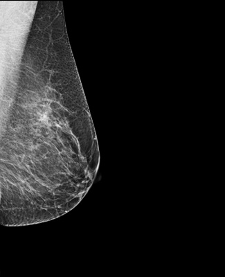
X-rays are a form of ionising radiation which penetrates the body to form pictures on film or digital imaging (eg. a computer screen). This type of radiation is used widely in medical facilities to perform diagnostic imaging procedures.
X-rays provide valuable information about the inside of your body and are important in helping your doctor make an accurate diagnosis.
Diagnostic imaging with no radiation
Other forms of diagnostic imaging such as magnetic resonance imaging (MRI) and ultrasound are procedures that do not expose you to ionising radiation.
Radiation exposure
Studies have linked high doses of ionising radiation (>50mSv) to an increased risk of cancer. Living cells can be damaged by X-rays. However, the risk is extremely low with the radiation dose you will receive from a mammogram.
Table 1 compares the radiation dose of a screening mammogram, (which involves 2 X-rays of each breast), to other imaging tests.
| Radiology |
Typical effective |
Equivalent period of natural background radiation |
|---|---|---|
|
X-ray Examination |
||
|
Chest |
0.02 |
5 days |
|
Pelvis |
0.6 |
5 months |
|
Mammogram |
0.7 |
24 weeks |
|
Lumbar spine |
1.5 |
12 months |
|
CT Examination |
||
|
Head |
2.0 |
18 months |
|
Chest |
7.0 |
5 years |
|
Abdomen |
8.0 |
5 years |
Screening mammography and radiation
A screening mammogram is an X-ray of the breast tissue for women without any breast symptoms. It uses low doses of radiation (about 0.7mSv for 4 X-rays).
Breast tomosynthesis
Also known as 3D mammography, tomosynthesis uses special computer software to create a 3D image using X-rays taken at different angles. The radiation dose is slightly higher than the dosage used in screening mammography (~25%).
X-ray safety
X-rays are produced only when a switch is momentarily turned on, like a light switch. No radiation remains after the switch is turned off.
BreastScreen WA contracts licensed medical physicists (a scientist specialising in physics in medicine) to routinely evaluate the radiation dose from X-ray equipment used to ensure it is as low as possible and does not exceed regulatory limits.
Factors affecting X-ray dose
The radiation dose from a screening mammogram is the amount of X-rays that are absorbed in the breast tissue. Two of the major factors affecting radiation dose are the amount of compression and the thickness and structure of the breast.
Breast compression during a screening mammogram reduces the radiation dose significantly since a thinner amount of breast tissue absorbs less radiation. It also separates overlapping folds of breast tissue that may obscure small abnormalities.
Breast implants can block a clear view of the breast tissue making mammograms less effective in breast cancer detection.
Therefore, radiation exposure will be greater in women with implants, because more X-rays need to be taken (between 6 - 8) and less compression is used.
Risks and benefits
Risks
The risk of getting cancer from a screening mammogram is considered to be very low.
Table 2 below shows the additional risk in a person’s lifetime of developing cancer from X-ray procedures at each examination.
| Examination | Lifetime additional risk of cancer per exam* |
|---|---|
| Negligible Risk | |
| Chest, teeth, arms & legs, hands & feet x-rays | Less than 1 in 1,000,000 |
|
|
Minimal Risk |
| Skull, head, neck x-rays | 1 in 1,000,000 to 1 in 100,000 |
| Very Low Risk | |
|
Hip, spine, abdomen, pelvis x-rays, CT head, breast mammography |
1 in 100,000 to |
| Low Risk | |
|
Kidney & bladder [IVU], Stomach – barium meal, CT chest, CT abdomen |
1 in 10,000 to 1 in 1,000 |
Table 2 Source: Adapted from A guide for Medical Imaging. Australian Radiation Protection and Nuclear Safety Agency, 2015.
*These risk levels represent very small additions to the 1 in 3 chance we all have of getting cancer throughout our life time.
If you are pregnant, it is advisable you postpone your screening mammogram until after the birth. A screening mammogram is not recommended for women who suspect they are pregnant.
Benefits
The low dose of radiation used in a screening mammogram has not been proven to cause harmful effects. The benefit of early diagnosis and treatment of breast cancer far outweighs the risk of the small amount of radiation received during a screening mammogram.
How often should I have a mammogram?
At least every two years unless there are reasons for more frequent mammograms. If breast cancer is detected early, there is a good chance it can be treated successfully.
Book online or phone 13 20 50
When making your appointment, please let us know if you:
- have breast implants
- require an interpreter
- use a wheelchair.
These things may make your appointment a little longer.
If you need an interpreter, please call the Translating and Interpreting Service (TIS) first on 13 14 50 and ask to be connected to the BreastScreen WA call centre on 13 20 50.
NOTE:
Wheelchair access is available at all BreastScreen WA services.
Appointments are available 7:30am-5:45pm on weekdays and 8:15am-11:30am on Saturdays at most BreastScreen WA clinics.
- Please bring your Medicare card with you to your appointment.
- Women whose breasts become tender before their periods find it more comfortable to have a mammogram during or just after a period.
- Please don’t wear talcum powder or deodorant on the day of your appointment. It may show on the X-ray picture.
- Please be aware you will be reading information and signing a consent form at the appointment.
Get to know your breasts and what is normal for you. Look in the mirror at your breasts and feel your breasts from time to time.
If you notice any unusual changes in your breasts such as lumps, nipple discharge, or persistent new breast pain, even if your last screening mammogram was normal, please see your GP promptly.
Ask your GP about breast health at your next check-up.
Use our contact form or call BreastScreen WA on:
13 20 50 for appointments
9323 6700 for information
BreastScreen WA has metropolitan, regional and mobile screening services.
There are twelve permanent clinics located in:
Albany, Bunbury, Cannington, Cockburn, David Jones - Perth City, East Perth - Mardalup, Joondalup, Mandurah, Midland, Mirrabooka, Padbury, and Rockingham.
Appointments are available 7:30am-5:45pm on weekdays and 8:15am-11:30am on Saturdays at most BreastScreen WA clinics.
Mobile breast screening services visit outer metropolitan areas and country towns every two years. Some towns are visited annually.

